 |
Neural
crest transplants Tim Carl |
GFP-labelled
crest transplant
|
|
|
|
|
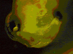
| Eggs
are obtained from hormone-stimulated female Xenopus laevis and
fertilized in vitro following established lab procedures. Fertilized eggs are dejellied using 2% cysteine (pH 8.0) and reared at room temperature in 10% Holtfreter's solution. Larvae are staged according to the normal table of Nieuwkoop & Faber (1967). In Xenopus
laevis, cranial neural crest migration begins in the late neurula (stage
19). Embryos are
placed in Petri dishes lined with 2% agar and Small segments
of the cranial neural folds from host embryos are ablated Care must be taken to remove the overlaying ectoderm. Grafts is held
in place initially with modeling clay and Specimens can be viewed using a stereo dissecting microscope equipped with epifluorescence optics. |
| |
| |