Klymkowsky Lab On-line Methods
wax-sectioning & staining
after Kelly et al 1989. Histological preparation of Xenopus laevis oocytes and embryos. in Meth. Cell Biol. 36:389.

Table of Methods updated - 14 September 2003
This section is being modified! - stay tuned

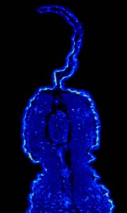
Plakoglobin in the epidermis


  1. Fix samplesin MEMFA and then into Dent's Fix (20% DMSO/80% Methanol)
  2. If specimens need to be bleached, bleach in Curis Bleach for 1/2 hour under white light. Store specimens at -20°C in 100% methanol
  3. place embryos into 100% ethanol (dehydrated with molecular sieve (type 3A - BioRad)
  4. infiltrate specimens with parafin -- place embryos into glass petri dish containing molten paraplast (58°-59°C). Repeat with fresh molten wax.
  5. fill a base mold with 5mm parafin -- move embryos into mold with pipette
  6. 2X washes with xylene, 15 minutes each
  7. Add 50% EtOH and 50% Hemo-De, 20 minutes for large specimens
  8. Replace with 100% wax, 2 hours at 58°C
  9. Replace with 100% wax, overnight at 58°C *All times are approximate.  Large samples require longer time. 
  10. section

  1. Heat steel embedding dish over flame briefly before adding wax  (this prevents bubbles from forming and allows even cooling of the wax)
  2. Place enough wax to fill embedding dish
  3. Add specimen to dish and orient into desired position
  4. Place embedding ring on the embedding dish (make sure that there is enough wax in the dish so that the embedding ring becomes slightly immersed in the wax - this holds the ring in place)
  5. Add hot wax to the dish until it nearly reaches the top of the embedding ring
  6. Carefully reorient the specimen if it has moved
  7. Cool on cool surface, overnight
  8. Remove dish and section

Curis bleach

Table of Methods