IF organization & functions in skeletal muscle |
Based on the apparent lack of function of IFs in cultured cells, we reasoned that IFs must function in an organismic context, most likely in the strengthening of cells against mechanical deformation. It was therefore relatively obvious to look for IF functions in muscle, since it generates its own mechanical stress. All types of muscle cells express the muscle-specific IF protein desmin. Desmin is a member of the vimentin-like IF protein group. In addition to desmin, other IF proteins are expressed during various stages of their development. Nestin and vimentin are often expressed early in myogenic differentiation, whereas synemin and paranemin are expressed later. In Xenopus, the first muscle to form is the dorsal myotomal muscle, which plays a role in the swimming movements of tadpoles. |
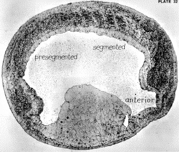
This dorsal myotome forms from the somites. Somites form in an anterior to posterior wave, with one forming ~ every 40 minutes. The cells that will form the somites are located on both sides of the anterior-posterior axis, and are originally oriented perpendicular to it. As somites form, these cells rotate ~90°, so as to be oriented parallel to the neural tube.
Image from Hausen & Riebesell.1991. The Early Development of Xenopus laevis. Springer Verlag. |
In contrast to the situation in the mouse and the chick, where the expression of vimentin precedes the expression of desmin during myogenic differentiation, vimentin is never expressed in the cells of the dorsal myotome . This is significant in that desmin, when it is first expressed, integrates into a preexisting vimentin filament network. |
Dent,
J.A., A.G. Polson & M.W. Klymkowsky. 1989. A
whole-mount immunocytochemical analysis of the expression of the intermediate
filament protein vimentin in Xenopus. Development 105: 61-74. |
In contrast, in Xenopus, desmin filament assembly appears to occur de novo. |
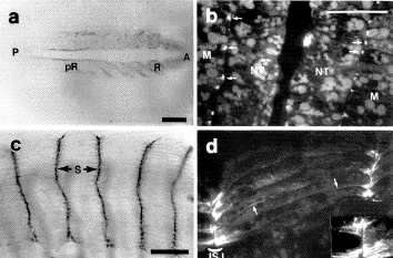
When first expressed, prior to somite segmentation/rotation (pR in panel a) desmin is found localized to the tips of the myogenic precursor cells. Following segmentation, desmin is concentrated at the ends of the myotomal cells. Cary, R.B. & M.W. Klymkowsky. 1994. Desmin organization during the differentiation of the dorsal myotome in Xenopus laevis. Differentiation, 56:31-38. |
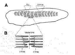
Only later in muscle differentiation does desmin come to be closely associated with the Z-lines of the muscle cell. Alongitudinal IF network, typical of vimentin-expressing myogenic cells, never appears. This appears to be specific property of desmin, if vimentin is expressed in these myotomal cells, it does form a longitudinal network. The difference between the two proteins appears to reside in the N-terminal, non-helical head domain. |
Cary,
R.B. & M.W. Klymkowsky. 1994. Differential
organization of desmin and vimentin in muscle is due to differences
in their head domains. J. Cell Biol., 126: 445-456. |
Schweitzer
S.C., M.W. Klymkowsky, R.M. Bellin, R.M.Robson, Y. Capetanaki & R.M.
2001. Paranemin
and the organization of desmin filament networks. J
Cell Sci. 114:1079-1089 |
In more recent studies, carried out in collaboration with Bob Evans, we found that the formation of extended desmin networks in mammalian cells was greatly enhanced by the presence of another IF protein, paranemin. |
We began our studies in on muscle IF function there was experimental evidence from studies on cultured cells that the integrity of the IF network was not required for the establishment of the contractile apparatus. |
We built on the work of Albers & Fuchs (1987) who found that truncated epidermal keratins acted as dominant negative mutations, disrupting keratin filament organizaton when expressed in epithelial cells. We constructed a similar truncated forms of vimentin and desmin. When DNA encoding these proteins was injected into fertilized Xenopus eggs, it was expressed in myotomal cells. |
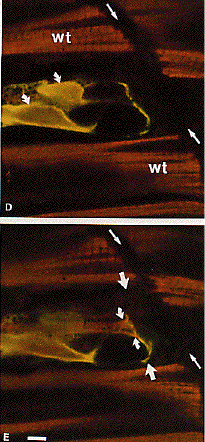
Using a combination of immunofluorescence, immunoelectron and confocal microscopy, we were able to show that these mutant IF proteins were able to disrupt desmin filament organization in myotomal cells. |
|
1953-2004
Michael Klymkowsky and associates last updated: 24 October 2004 |