| 12. Getting through membranes | Tweet |
|
||
|
|
|
|
Lipid bilayer membranes pose a serious barrier to the movement of larger molecules and small, hydrophilic molecules. This barrier property enables cells to use membranes as boundaries, to keep components inside and to exclude many unwanted molecules. |
|
The permeability barrier properties of the lipid portion of the membrane are rather non-specific. If you are hydrophilic, this barrier is quite high. On the other hand, if you are hydrophobic, the barrier is low, but you may find it difficult to leave the membrane once you enter it. The more hydrophobic a molecule, the more readily it passes into a membrane. The cell can control which types of molecules enter and leave using proteins embedded within the membrane - we will return to these shortly. The combination of lipids and proteins can produce quite complex behaviors. Some biological membranes act as batteries, storing electromechanical energy, others can carry signals over long distances (your nervous system uses membranes this way.) |
|
Membrane evolution: The modern cell membrane is composed of lipids with hydrocarbon or isoprene chains, typically ranging from 16 to 20 carbons in length.
The earliest membranes were likely to have been composed of similar, but simpler molecules with shorter hydrophobic chains. |
|
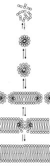
Based on the properties of lipids, we can map out a plausible sequence for the appearance of membranes. Lipids with very short hydrophobic chains, 2 to 4 carbons long, can dissolve in water (can you explain why?) As the lengths of the hydrophobic chains increases, the molecules begin to self-assemble into micelles. By the time the hydrophobic chains reach ~10 carbons in length, the molecules begin to associate into semi-stable bilayers. Bilayers can "capture" regions of water. Bilayer stability increases further as hydrophobic chain length increases. At the same time, membrane permeability decreases. It is a reasonable assumption that the earliest biological systems used shorter chain lipids to build their "proto-membranes" and that these membranes were relatively leaky. [link] |
|
|
Cells must exchange materials with the outside world in order to maintain their structure, to grow and to reproduce. The appearance of more complex lipids, capable of forming more impermeable membranes must therefore have depended upon the appearance of mechanisms that enabled hydrophilic molecules to pass through membranes. The process of interdependence of change is known as co-evolution. |
|
Note: do you understand that this video represents a three-dimensional process? |
|
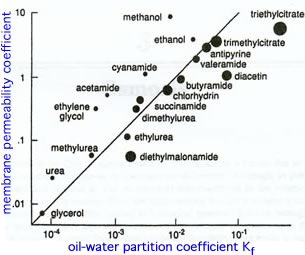
Aqueous pores in membranes: In the late 1940's Collander and colleagues studied the movement of molecules into cells. |
|
|
They noticed that small, water soluble molecules entered cells faster than predicted based on the assumption that the membrane acted like a simple hydrophobic barrier - this assumption was known as Overton's Law. Collander et al., postulated that membranes contained features that enabled them to act as molecular sieves. These "features" turned out to be protein pores, channels,and pumpswhich we will discuss further on. |
|
Diffusion: A group of particles, initially confined to a small volume will, over time, disperse. This movement is produced by collisions with neighboring molecules, which are in constant motion. This asymmetry of behavior is the basis of the second law of thermodynamics, it is described by the concept of entropy. |
|
If there is a difference between the concentration of a molecule in one region compared to another, that is known as a concentration gradient. There will be a netflux from the region of high concentration to the region of low concentration. Both solute and solvent can diffuse. Osmosis is the net movement of solvent. |
|
|
Cells are packed full of molecules. These molecules take up space, space no longer occupied by water. It is therefore common for the concentration of water outside of the cell [H2O]out to be higher than the concentration of water inside the cell [H2O]in.
|
|
|
|
The water gradient is capable of doing work -- it can lift a fraction of the solution against the force of gravity. At equilibrium, the force generated by osmosis is balanced by the weight of the levitated solution. In an important sense, the concentrated cytoplasmic organization of the cell represents stored energy. |
|
|
|
Dealing with osmosis: The water concentration gradient across the plasma membrane of most organisms leads to an influx of water into the cell, particularly if there are water channels [aquaporins] in the membrane. As water enters the cell, the plasma membrane will expand. If the influx of water continues, the membrane will burst like an over-inflated balloon. Organisms, such as plants, fungi and bacteria, use cell walls to deal with the influx of water. The cell wall is a specialized and relatively rigid polymer-based, extracellular matrix located outside of the plasma membrane. The space between the cell wall and the plasma membrane is known as the periplasmic space. As water enters the cell (by osmosis), the plasma membrane is pressed up against the cell wall at the expense of the periplasmic space. The force exerted by the cell wall on the membrane balances the force of water entering the cell. |
|
|
When the two forces are equal, the net influx of water into the cell stops. When the [water] outside decreases, this pressure is reduced and the plant wilts. This is a passive effect, built into the wall when it was first assembled. Once the cell wall has been built, a cell with a cell wall does not need to expend energy to resist osmotic effects. |
|
|
Dealing with osmosis without a cell wall: Animal cells do not have a rigid cell wall. This allows them to be active predators, moving rapidly and engulfing their prey (organisms with a rigid cell wall can't do that!) It also means, however, that they must use other mechanisms to deal with the effects of osmosis. |
|
Most free living protozoa, single celled eukarya without a rigid external wall, live in dilute aqueous solutions, where osmotic effects are significant. They deal with the constant in-flux of water by actively pumping the water that flows in back out again using an organelle known as the contractile vacuole. Water accumulates within the contractile vacuole, a membrane-bounded structure, which inflates. |
 |
|
To expel the water, the vacuole connects with the plasma membrane and is squeezed by cytoskeletal systems within the cytoplasm. This squirts the water out of the cell. The process of vacuole contraction is an active one, it involves work and requires energy. |
|
|
|
Questions to answer |
|
|
Questions to ponder |
|
|
|
| replace with revised beSocratic activity |
|
|
modified 10-May-2014
|
|