Intermediate filament functions: dismissed & revealed
topics : wnt signaling | sox networks | heart induction | EMT & apoptosis

In the early 1980's, when I started working on IFs, they were arguably the most enigmatic component of the cytoskeleton.   While microtubules and microfilaments are ubiquitous and essential components of eukaryotic cells, IFs were clearly not. 

In fact there is an evolutionary oddity, insects (such a Drosophila melanogaster) appear not to have cytoplasmic IFs, although they do have lamins. 

Nevertheless, IFs are a major component of the cytoskeleton in many cell types and a nuclear lamina appears to be present in most eukaryotes.

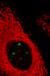
Confocal image of the keratin-type IF network (red filaments) of a cultured epithelial cell. The cell also expressed a nuclealy-localized green fluorescent protein chimera (plakophilin-1-GFP).

The typical approach to studying the function of a structure is to disrupt it, and then watch the effects on the cell.
 
A major obstacle to understanding IFs was the absence of reagents that specifically disrupted IF organization.

It was in this light that the discovery that the injection of anti-IF antibodies can specifically disrupt IF network organization was of significance.

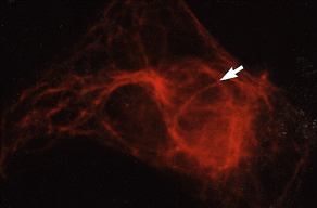
Injection of antiIFA into mouse fibroblasts induces the collapse of the IF network. Sometimes, the collapsed IFs can be seen to influence the organization of microtubules (roll over -vimentin is green, microtubules are red)


Antibody injection can be used to disrupt IF organization in a wide range of cells.

Working initially at Cold Spring Harbor, and later in London, E. Birgitte Lane generated a number of monoclonal antibodies directed against the keratin-type IF proteins of simple epithelial cells.

Different monoclonal antibodies had different effects on keratin filament organization.

IFs had been postulated to play a major roll in the establishment and maintanence of cellular morphology. We used antibody injection to test these ideas.

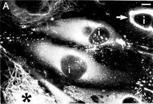
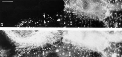
Injection of the monoclonal antikeratin antibody LE61 into cultured epithelial cells induces the disruption of the keratin-type IF network.

One can disrupt keratin organization in one cell and examine desmosome integrity and keratin organization in neighboring cells.

The surprising result was that there was no apparent change in cellular morphology, movement or intracellular organization.


This basic observation has been confirmed by a number of labs through the analysis of cell variants that completely lack cytoplasmic IFs and through the use of dominant negative mutant versions of IF protein.

In particular our long-time collaborator Bob Evans carried out an elegant and extensive analysis of variants of the human cell line SW13 that lacked all detectable cytoplasmic IFs, and found no apparent difference compared to sister lines that contained IFs.


Klymkowsky, M. W (1981). Intermediate filaments in 3T3 cells collapse after the intracellular injection of a monoclonal anti-intermediate filament antibody. Nature 291, 249-251

Klymkowsky, M. W (1982). Vimentin and keratin intermediate filament systems in cultured PtK2epithelial cells are interrelated. EMBO J 1, 161-165

Lane, E.B. & M.W. Klymkowsky. 1982. Epithelial tonofilaments: investigating their form and function using monoclonal antibodies. Cold Spring Harbor Symp. Quant. Biol. 46:387-402.

Klymkowsky, M. W., Miller, R. H. & Lane, E. B (1983). Morphology, behavior, and interaction of cultured epithelial cells after the antibody-induced disruption of keratin filament organization. J. Cell Biol 96, 494-509

Klymkowsky, M. W., Bachant, J. B. & Domingo, A (1989). Functions of intermediate filaments. Cell Motil. Cytoskel 14, 309-331

Klymkowsky, M.W. & R.M. Evans. (1995). Intermediate filaments in human pathology.
in Principals of Medical Biology.  E.E. Bittar and N. Bittar, editors. JAI Press, Inc. pp. 147-186.


1953-2004 Michael Klymkowsky and associates
last updated:24 October 2004
||