Looking to the organism for IF functions |
The lack of an overt and dramatic phenotypes following disruption of IF organization in culture cells lead a number of labs, including ourselves, to transfer our work from cultured cells to intact organisms. The real breakthrough in this area was made by Elaine Fuchs & colleagues, who found that the IF networks of epidermal cells were essential for the mechanical integrity of skin. Similar conclusions were derived from studies E.B. Lane and E. Epstein studying human epidermolytic diseases . Since then, it has been well established that a large number of epidermolytic (fragile-skin) diseases of humans are due to mutations in epidermal IF proteins. |
More recently, mutations in the muscle-specific IF protein desmin and nuclear IF-like lamin A/C proteins have have been linked to both cardiomyopathy, and in the case of lamin lipodsystrophy. |
Looking into Xenopus Beginning in about 1986, I turned to studying the organization and function of IFs in the clawed frog Xenopus laevis. We choose Xenopus because of the speed at which experiments could be performed and its low cost as an experimental system. One of first tasks was to develop antibodies that could be used to visualize IFs in Xenopus. Using insoluble residues of Xenopus A6 cells as an antigen, we generated monoclonal antibodies directed against nuclear lamins (14a9), vimentin (14h7) and keratin (1h5). these (together the anti-tubulin antibody E7) are availalbe from the Developmental Studies Hybridoma Bank. |
|
|
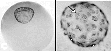
Our first studies were on the organization of the keratin filament network present in the oocyte and early embryo. We used whole-mount immunocytochemistry with the monclonal antikeratin antibody 1h5 to characterize the keratin filaments network of the late stage and maturing oocyte, egg and early embryo |
These studies have been beautifully
extended by Dave
Gard & colleagues at the using sections and confocal imaging.
The keratin network of the late stage oocyte is asymmetrically organized, with a distinct animal-vegetal asymmetry. That this asymmetry is an inherent property of the oocyte cytoplasm was revealed through experiments in which mRNA encoding Xenopus vimentin was injected into either the oocyte's animal or vegetal hemisphere. Only punctate structures were formed in the animal cortex, while long filaments were assembled in the vegetal cortex. |
Dent,
J.A., R.B. Cary, J.B. Bachant, A. Domingo & M.W. Klymkowsky. 1992. Host
cell factors controlling vimentin organization in the Xenopus oocyte. J.
Cell Biol.119: 855-866. |
Normally highly insoluble, the
keratin network of the oocyte is completely disassembled into soluble monomers
using meiotic maturation. A cartoon of keratin organization and reorganization during oocyte maturation and following fertilization. |
Klymkowsky, M.W., L.M. Maynell & A.G. Polson. 1987. Polar asymmetry in the organization of the cortical cytokeratin system of Xenopus laevis oocytes & embryos. Development100: 543-557. |
The surprising observation here was the apparently disassembly of the keratin network as the oocyte re-entered active meiosis and prepared for fertilization. Biochemical studies, carried out by Jeff Bachant revealed that the keratin network was completely disassembled into soluble oligomers that appeared larger than the expected tetramers. |
Bachant,
J.B. & M.W. Klymkowsky. 1996.
A non-tetrameric species is the major soluble form of keratin in Xenopus
oocytes and rabbit reticulocyte lysates. J. Cell Biol., 132: 153-165. |
|
Keratins and gastrulation defects |
1953-2004
Michael Klymkowsky and associates last updated: 7 April 2004 |